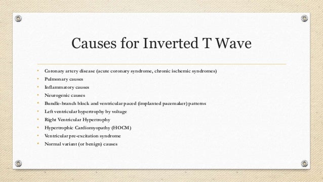
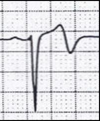
Inverted T Wave On Ecg Causes
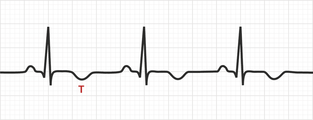
The T Wave Physiology Variants And Ecg Features Ecg Echo
T Wave Litfl Medical Blog Ecg Library Basics
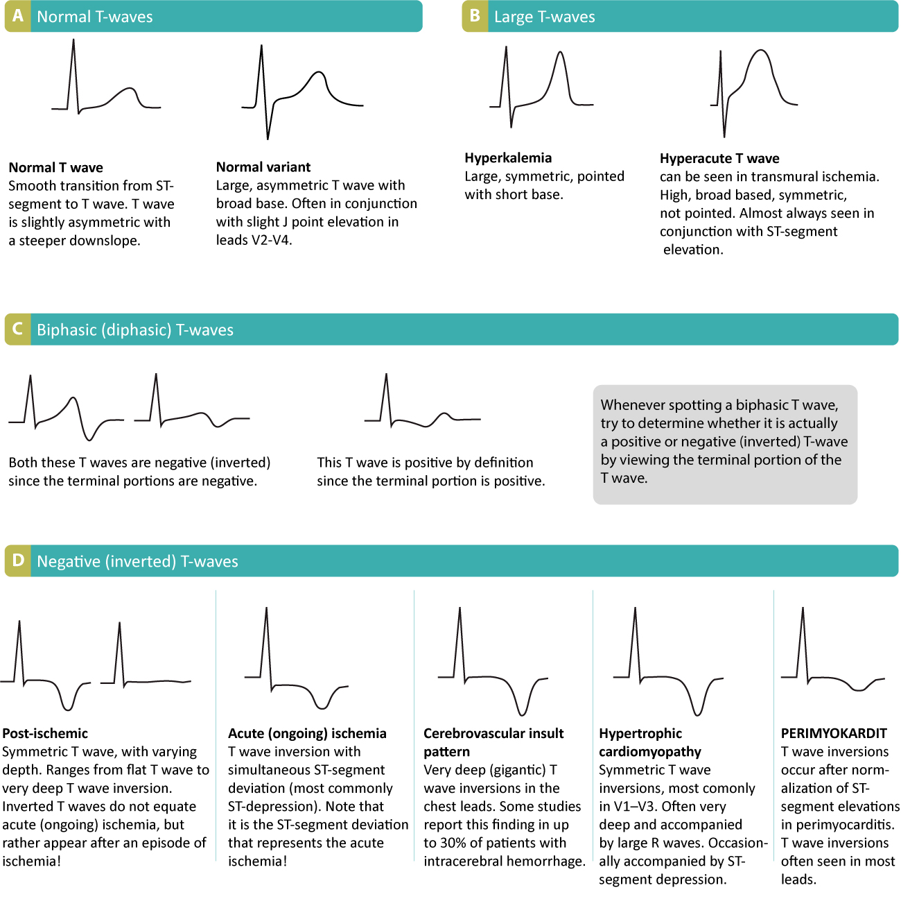
T Waves In Ischemia Hyperacute Inverted Negative Wellen S Sign De Winter S Sign Ecg Echo
T Wave Wikipedia

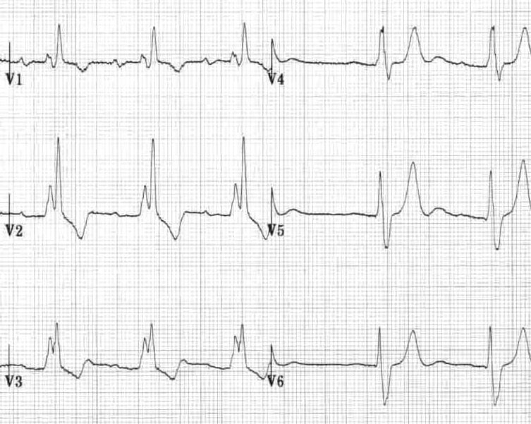
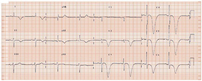
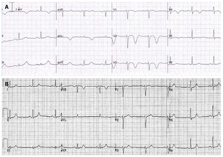
T Wave Inversion In Leads V1 V6 In A 38 Year Old Symptomatic An Download Scientific Diagram

Causes Of T Wave Inversion In Ecg Cardiology And Ccu Facebook

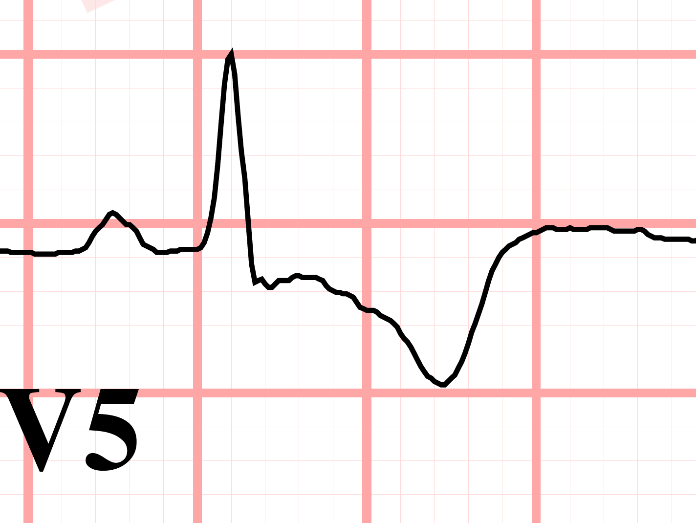
Inverted in lead avr.
Inverted t wave on ecg causes.
Lesson Title The T Wave
What Is T Wave Inversion Quora
Deep T Wave Inversion Thoracic Key

Mechanism Of Ischemic T Wave Inversion Youtube
Ecg T Wave Inversion Dr Malala Rajapaksha Cardiology Unit Genera

Dr Smith S Ecg Blog Reversible T Wave Inversion It Reverses Then Evolves Then Reverses When Ischemia Is Gone Normalization Of T Waves Not Pseudonormalization

St Depression T Wave Inversion Causes Pathophys Diagnosis Cardiology Medstudent Ekg

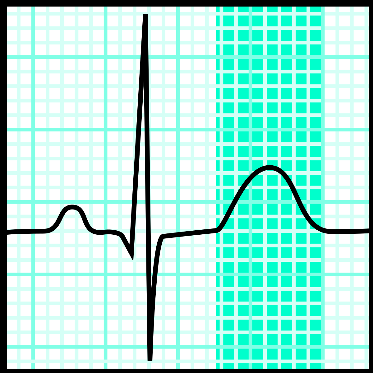
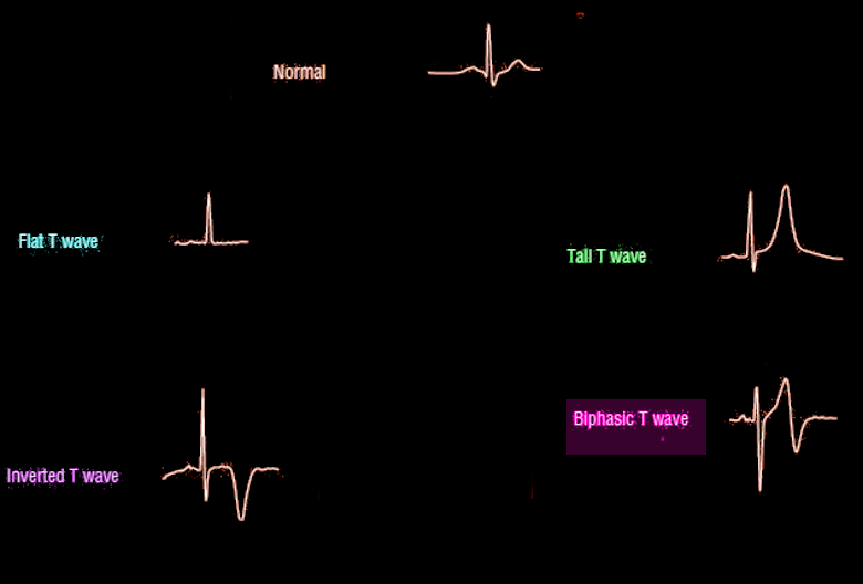
Ecg Normal And Inverted T Wave Waves Normal Ekg
Basic Electrocardiography Guide To Diagnostic Tests
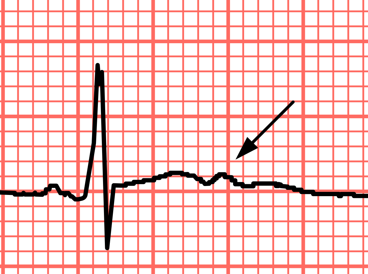
U Wave Wikipedia
The Inverted T Wave Differential Diagnosis In The Adult Patient

68 Causes Of T Wave St Segment Abnormalities Learntheheart Com

Chest Pain With Diffuse T Wave Inversion Photo Quiz American Family Physician

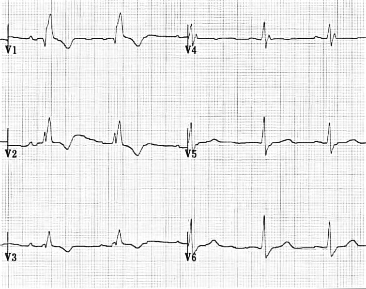
Resting 12 Lead Ekg Showing Symmetric T Wave Inversion In Right Download Scientific Diagram
T Wave Inversion Test Findings Medschool

Ecg In A Patient With Arvd C Epsilon Waves And Inverted T Waves Download Scientific Diagram
Cardiac And Non Cardiac Causes Of T Wave Inversion In The Precordial Leads In Adult Subjects A Dutch Case Series And Review Of The Literature
Right Bundle Branch Block Rbbb Litfl Ecg Library Diagnosis
Https Encrypted Tbn0 Gstatic Com Images Q Tbn 3aand9gcrpzmybk 8i8errcymlgjsala2cz97lgve 7noquyscvllzqdr2 Usqp Cau
St Segment Depression In Myocardial Ischemia And Differential Diagnoses Ecg Echo
Emdocs Net Emergency Medicine Educationecg Pointers Intracranial Hemorrhage Emdocs Net Emergency Medicine Education

Dr Smith S Ecg Blog Chest Pressure During Exertion Evolution Of Inverted T Waves And Troponins Surprise Angiogram
Ecg T Wave Article Statpearls

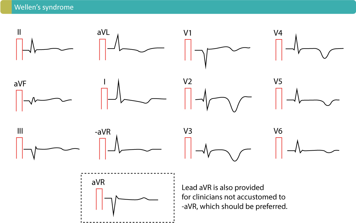
Wellens Syndrome Litfl Medical Blog Ecg Library Eponym
Source : pinterest.com
